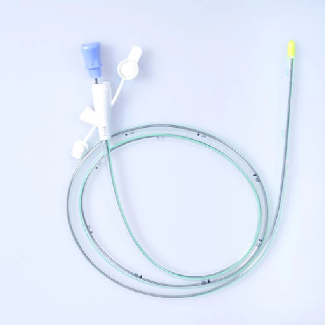


Triple Lumen Nasogastric Tube with Temperature and Pressure Sensors
Release time: 2026-09-15
From "Passive Channel" to "Active Monitor": A Paradigm Shift
Nasogastric tubes have been used in clinical practice for over a century. Their basic functions have always been clear: gastrointestinal decompression, enteral nutrition delivery, and gastric content sampling. As a "passive channel," the tube generates no data, contributes nothing to decision-making, and provides no feedback on patient status. But for critically ill patients receiving mechanical ventilation, relying on a nasogastric tube alone for nutritional support is becoming insufficient. The reason lies in a long-standing clinical challenge: the immeasurability of transpulmonary pressure.
Transpulmonary pressure is the difference between alveolar pressure and pleural pressure, and is considered the mechanical parameter most closely associated with ventilator-induced lung injury. When a patient has spontaneous breathing, airway plateau pressure alone cannot accurately assess the actual stress experienced by lung tissue, because the contribution of pleural pressure is ignored. Pleural pressure cannot be measured directly. The alternative used in clinical practice is esophageal pressure monitoring—at the lower third of the esophagus, where the esophageal wall is separated from the pleura by only a thin layer of connective tissue, the pressure within the esophageal lumen can accurately reflect pleural pressure. Based on synchronized esophageal and airway pressure data, transpulmonary pressure can be calculated in real time.
The problem is that obtaining esophageal pressure requires a dedicated esophageal manometry catheter, typically a balloon catheter. This means that a mechanically ventilated patient who needs enteral nutrition and also requires esophageal pressure monitoring must have two tubes inserted—one for feeding and one for pressure measurement. This is the logical starting point for the "sensing nasogastric tube": why not let one tube do both?
Technical Implementation: How Sensors Enter the Tube Wall
Integrating a pressure sensor into the wall of a nasogastric tube presents a core challenge: balancing size and precision.
Traditional esophageal pressure monitoring relies on an inflatable balloon as a pressure transmission medium. Once inflated, the balloon conforms to the esophageal wall and transmits pressure to an external sensor. The problems with this approach are that the balloon inflation volume must be precisely titrated—too much or too little affects reading accuracy; the balloon position must be repeatedly verified; and changes in patient position can alter the conformity between the balloon and the esophagus.
The emergence of micro-MEMS pressure sensors offers another path. When sensor packaging can be reduced to the 0.3-millimeter scale, it can be embedded directly into the tube wall without needing a balloon as an intermediary. The sensor contacts the esophageal wall directly and outputs an electrical signal rather than pneumatic pressure, achieving sampling frequency and response speed far beyond balloon systems. More critically, this design circumvents a key patent barrier. Chinese patent CN113766873A protects a "sensor plus close-range inflatable balloon" combination. Any esophageal manometry catheter using a balloon as a physical separator may fall within its scope of protection. A micro-sensor design embedded directly into the tube wall bypasses the "balloon" as an essential element from a technical standpoint.
Clinical Data Support: The Evolution of Evidence for Transpulmonary Pressure Monitoring
The clinical value of esophageal pressure monitoring is not a new concept, but the evidence base has undergone significant evolution over the past fifteen years.
The EPVent study, published in 2008, was a milestone in this field. The study enrolled 61 patients with acute lung injury and ARDS with a PaO₂/FiO₂ ratio below 300 mmHg, comparing esophageal pressure–guided PEEP settings with a low-PEEP/FiO₂ table strategy. The results showed that the esophageal pressure–guided group had better oxygenation indices (280 mmHg vs. 191 mmHg) and lung compliance (45 vs. 35 mL/cmH₂O), with higher PEEP settings (17 vs. 10 cmH₂O) and end-expiratory transpulmonary pressure maintained at a more reasonable level (0.1 vs. −2 cmH₂O). The significance of the latter figure is that under the low-PEEP/FiO₂ table strategy, end-expiratory transpulmonary pressure was negative, meaning the alveoli had already collapsed at end-expiration.
The EPVent-2 study, published in 2019, presented more complex results. This multicenter randomized controlled trial enrolled 200 patients with moderate-to-severe ARDS, comparing an esophageal pressure–guided strategy with a high-PEEP/FiO₂ table strategy. The study failed to demonstrate a significant advantage of esophageal pressure guidance on the primary outcome, but post-hoc analysis suggested lower mortality among patients whose end-expiratory transpulmonary pressure was maintained within the range of −2 to 2 cmH₂O.
The 2024 updated monitoring guidelines recommend that directly measured end-expiratory transpulmonary pressure should be titrated between 0 and 2 cmH₂O to avoid alveolar collapse in dependent lung regions, while end-inspiratory transpulmonary pressure should be kept below 20 cmH₂O. These recommended values are being progressively refined, reflecting a shift in clinical understanding of transpulmonary pressure targets from "higher is better" to "precise range."
From Weaning Assessment to Nutrition Safety: The Multiple Roles of a Single Tube
The applications of esophageal pressure monitoring extend beyond PEEP titration.
In weaning assessment, esophageal pressure provides information that conventional spontaneous breathing trials cannot capture. A spontaneous breathing trial judges weaning timing by observing whether a patient can tolerate spontaneous breathing, but passing the trial does not equate to successful weaning. The EXTEND ARDS-J study, launched in 2024, is testing a hypothesis: whether the magnitude of esophageal pressure changes during a spontaneous breathing trial can predict trial failure and the risk of reintubation within 48 hours after extubation. The background of this study is that excessive inspiratory effort in ARDS patients may induce pendelluft phenomenon, leading to injury of dorsal lung regions in the supine position.
On the nutrition management side, a tube with intragastric pressure monitoring capability provides an additional safety dimension. Gastric residual volume monitoring is a traditional means of assessing enteral nutrition tolerance, but the aspiration method alone has accuracy limitations. Combining intragastric pressure data with gastric residual volume trends allows for a more comprehensive assessment of gastric emptying function. Continuous monitoring of intra-abdominal pressure helps with early identification of intra-abdominal hypertension, which has early-warning value in scenarios such as severe pancreatitis and post-abdominal surgery.
Engineering Perspective: The Distance from Laboratory to Production Line
Translating the above physiological principles into a mass-producible product involves several engineering decisions.
Sensor layout. The esophageal sensor must be positioned at the lower third of the esophagus to accurately reflect pleural pressure, while the intragastric sensor must be positioned below the diaphragm to obtain intragastric pressure readings. The spacing between the two sensor segments is determined by catheter insertion depth and patient anatomy, requiring a certain tolerance range.
Packaging protection. The sensors come into direct contact with the esophageal mucosa and gastric fluid, so the packaging material must meet biocompatibility requirements while resisting chemical corrosion from digestive fluids. A 14-day saline immersion test is the basic threshold for verifying long-term stability.
Signal integration. Multiple sensor signals need to be routed within the tube wall and integrated into a standard digital interface at the external end. The I²C protocol is a reasonable choice due to its simplicity and broad compatibility, but the flexibility and interference resistance of the wiring need to be verified in actual use.
Conclusion
Integrating pressure sensing into a nasogastric tube is not, at the technical level, a disruptive innovation. It is more like reassembling already mature components—micro-MEMS sensors, flexible circuits, standard digital interfaces—into an existing clinical tool.
But the significance of this "combinatorial innovation" should not be underestimated. It transforms esophageal pressure monitoring from a "separate procedure" requiring a dedicated catheter into data collection that is "completed along the way" during enteral nutrition support. For ICU patients who already have a nasogastric tube in place, this means transpulmonary pressure data can be continuously obtained without any additional invasive procedure.
When a single tube simultaneously serves the triple roles of feeding, decompression, and respiratory mechanics monitoring, the data density of intensive care is substantially enhanced. This enhancement requires no new beds, no new equipment, and no new staffing—only placing a sensor into the wall of a tube that is already there.